|
|
Plasma Membrane
- General function in cell containment
- Composition of membranes
- Lipid components
- Phospholipids (forms bilayer)
- Steroids, cholesterol (provide more fluidity)
- Waxes (provide more stability)
- Protein components: receptors, channels, transport proteins
- Fluid mosaic model: the fluid mosaic model basically describes the membrane as protein boats floating in a sea of lipids.
- Membrane dynamics
- Invaginates to take things in: phagocytosis, pinocytosis, endocytosis
- Buds off: exocytosis
- Changes shape: chemotaxis with the aid of changes in cytoskeleton
- Let's things through: small nonpolar molecules, others by the aid of channels or pumps
- Solute transport across membranes
- Thermodynamic considerations
- Mixing a charged ion with hydrophobilic lipid bilayer is thermodynamically unfavorable by entropy
- Therefore, ions can't pass through the lipid bilayer without assistance from channels/pumps
- Osmosis: water diffuses freely across the membrane, but not ions. So osmosis occurs readily
- Colligative properties depend on the number of solute dissolved in solvent
- Solvent will move as to dilute the component with more solute dissolved (given a semipermeable membrane that lets solvent pass but not the solutes)
- Osmotic pressure = pressure exerted by solvent via osmosis = counter pressure needed to prevent osmosis
- The bigger the difference in solute concentration, the bigger the osmotic pressure
- Too much osmotic pressure, and the cell will lyse (eg: if you put animal cells in water)
- Passive and active transport: things that can't readily diffuse across the membrane are transported across the membrane either without energy (passive) or with energy (active).
- Passive transport = no ATP needed = channels, facilitated diffusion = direction of movement is down a concentration gradient
- Active transport = ATP needed = sodium/ptassium pump = up/against a concentration gradient
- Sodium-potassium pump: 3 sodium (NA+) out, 2 potassium (K+) in. Thus, the cell maintains a negative resting potential.
- Membrane channels: lets ions through the cell membrane
- Membrane potential: the resting potential of the cell membrane is negative because of the sodium-potassium pump.
- Membrane receptors, cell signaling pathways, second messengers
- Many hormones can't cross the plasma membrane, so they bind to membrane receptors on the outside.
- Receptor binding triggers the production of second messengers.
- Second messengers cause a change inside the cell (through a protein kinase cascade).
- Cell signaling pathways:
- Contact signaling = physical contact triggers a change inside cell.
- Chemical signaling = chemical binding to receptor triggers a change inside cell.
- Nerves use neurotransmitters.
- The endocrine system use hormones.
- Electrical signaling = change in membrane potential triggers change in cell.
- Action potential along neurons propagates and cause release of neurotransmitters into synapse..
- Action potential along muscle cell membrane causes contraction.
- Exocytosis and endocytosis: exo = getting stuff out, endo = taking stuff in.
- Intercellular junctions
- gap junctions: connects two cells, and allows stuff to flow through between the cells.
- tight junctions: stitches/glues two cells together, and does not allow stuff to flow through between the cells. A series of cells with tight junctions also effectively forms an impermeable barrier.
- desmosomes: connects two cells together by linking their cytoskeleton. They are organized for mechanical strength, not an impermeable barrier.

Membrane-Bound Organelles and Defining Characteristics of Eukaryotic Cells
- Defining characteristics (membrane bound nucleus, presence of organelles, mitotic division)
- Defining characteristics = what sets eukaryotes apart from prokaryotes.
- Eukaryotes have a true nucleus (membrane-bound), while prokaryotes don't.
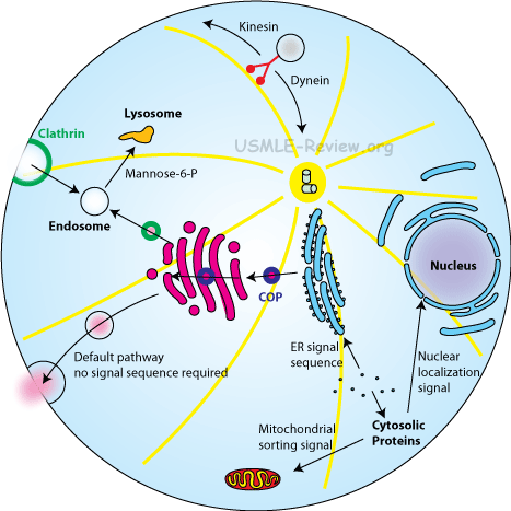
- Eukaryotes have membrane-bound organelles (ER, Golgi, lysosomes, mitochondria), prokaryotes don't.
- Eukaryotes divide by mitosis (all them chromosomes line up and stuff), prokaryotes undergo binary fission (no chromosomes, just a circular ring of DNA, no need for complex mitosis)
- Nucleus (compartmentalization, storage of genetic information)
- compartmentalization: nuclear membrane / nuclear envelope surrounds the nucleus.
- genetic information is stored inside the nucleus as DNA.
- Nucleolus (location and function)
- location is a region inside the nucleus.
- function is to transcribe ribosomal RNA (rRNA).
- Nuclear envelope, nuclear pores
- nuclear envelope is a double membrane system made of an outer and an inner membrane. Also called nuclear membrane.
- nuclear pores are holes in the nuclear envelope where things can pass into and out of the nucleus. Transcription occurs in the nucleus, and those transcribed RNA need to pass out of the nucleus. Things like transcription factors need to pass into the nucleus where they can access the DNA to be transcribed.
Membrane-bound Organelles
- Mitochondria
- site of ATP production: an apparatus called the ATP synthase makes ATP from ADP by utilizing the proton gradient as the driving force. The proton gradient is where the proton H+ concentration is higher in the inter-membrane space than the matrix of the mitochondria.
- self-replication; have own DNA and ribosomes.
- mitochondria replicate independently from the cell containing the mitochondria.
- mitochondria does not share the same genome with its host.
- mitochondria has their own ribosomes, which are different from the host's ribosomes in both sequence and structure.
- All these serve to support the endosymbiosis theory.
- inner and outer membrane
- Inner membrane surrounds the matrix.
- The folds of the inner membrane make up the cristae.
- Between the outer and inner membrane is the intermembrane space.
- The intermembrane space is high in protons H+.
- The outer membrane separates the mitochondria from the cytoplasm.
- Lysosomes (vesicle containing hydrolytic enzymes)
- Digests things like food and viral/bacterial particles.
- Things you want to digest gets into a vacuole by endocytosis or phagocytosis, and then the vacuole fuses with the lysosome. Anything inside gets digested by the hydrolytic enzymes.
- Endoplasmic reticulum:
- rough (RER) and smooth (SER)
- rough ER has ribosomes studded over it, smooth ERs don't.
- RER deals with protein synthesis, folding, modification, and export.
- SER deals with biosynthesis of lipids and steroids, and metabolism of carbohydrates and drugs.
- In the muscles, the SER or SR stores and regulates calcium.
- RER (site of ribosomes): the ribosomes attach to the outside of rough ER and synthesis protein into the lumen.
- role in membrane biosynthesis: SER (lipids), RER (transmembrane proteins)
- SER = makes lipids of the plasma membrane.
- RER = makes transmembrane proteins, carries them on its membrane, RER membrane forms vesicles and bud off, fuses with the plasma membrane, transmembrane proteins now on the plasma membrane.
- RER (role in biosynthesis of transmembrane and secreted proteins that cotranslationally targeted to RER by signal sequence)
- Transmembrane proteins, or proteins that are to be secreted (need RER vesicle) have a signal sequence right at the beginning.
- When ribosome starts making those proteins, they make the signal sequence first.
- Signal sequence recruits a signal recognition particle that drags it to the RER.
- ribosome now on the RER continues making the protein, but snakes it into the lumen.
- Signal sequence is clipped off.
- All ERs are connected to the nuclear membrane (an old aamc topic, no longer tested).
- Golgi apparatus (general structure; role in packaging, secretion, and modification of glycoprotein carbohydrates)
- looks like stacks of pancakes.
- modifies and/or secretes macromolecules for the cell.
- RER make protein → modified in the Golgi → buds off golgi and secreted out of cell by exocytosis.
- Glycoprotein = protein with attached saccharides.
- Golgi can glycosylate proteins as well as modifying existing glycosylations.
- Glycosylation affects protein's structure, function, and protect it from degradation.
- Peroxisomes: organelles that collect peroxides
- Contain oxidative enzymes for intracellular digestion
Cytoskeleton
- General function in cell support and movement
- Microfilaments (composition; role in cleavage and contractility)
- made of actin
- responsible for cytokinesis. Supports cell shape by bearing tension.
- Microtubules (composition; role in support and transport)
- made of tubulin
- responsible for mitotic spindle, cilia/flagella, intracellular transport of organelles and vesicles. Supports cell shape by bearing compression.
- Intermediate filaments (role in support)
- composition is varied.
- supports cell shape by bearing tension.
- Composition and function of eukaryotic cilia and flagella
- made of microtubules (eukaryotic)
- cilia can be for locomotion, sensory, or for sweeping mucus.
- flagella is used for locomotion.
- Centrioles, microtubule organizing centers. Microtubules radiate out of these barrel shaped structures, which are made of microtubules themselves.
Tissues Formed From Eukaryotic Cells
- Epithelial cells
- Formed from the ectoderm and endoderm
- Example: skin, intestinal epithelium
- Cancer of epithelial cells is called carcinoma
- Connective tissue cells
- Formed from the mesoderm
- Example: muscle, fat
- Cancer of connective tissue cells is called sarcoma
Cell Cycle and Mitosis

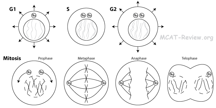
- Interphase and mitosis (prophase, metaphase, anaphase, telophase)
- Interphase
- G1 = Growth
- S = Synthesis (replicate DNA)
- G2 = Growth
- Prophase = Prepare (condense chromatin into chromosomes, break down nuclear membrane, assemble mitotic spindle, centriole pairs move toward opposite poles of the cell)
- Metaphase = Middle (Chromosomes line up in the middle)
- Anaphase = Apart (Sister chromatids pulled apart to opposite sides of cell)
- Telophase = Prophase in reverse = de-condense chromosomes, re-form nuclear membrane, break down mitotic spindle.
- Mitotic structures and processes
- centrioles, asters, spindles: responsible for pulling apart the sister chromatids
- chromatids, centromeres, kinetochores: sister chromatids are duplicated copies of the chromosome. chromatids are joined at the centromere. There's a protein at the centromere called the kinetochore, where spindle fibers attach to pull the chromatids apart.
- nuclear membrane breakdown and reorganization: for most eukaryotes, the nuclear membrane breaks down at the beginning of mitosis, and reforms at the end of mitosis around each of the two newly formed nuclei.
- mechanisms of chromosome movement: chromatids move apart during anaphase by the spindle fibers. Microtubules cause the chromosome movement.
- Phases of cell cycle: G0, G1, S, G2, M
- G0 = no more DNA replication or cell division. Examples include nerves and muscles.
- G1 = growth = make organelles, increase in cell size.
- S = DNA replication. Centrioles also replicated.
- G2 = growth = make organelles, increase in cell size.
- M = mitosis.
- Growth arrest: the cell cycle can be arrested for many reasons:
- Too much genomic mutation/damage causes a cell to arrest in M phase.
- Contact inhibition: normal epithelial cells stop growing when it gets crowded such that it's touching adjacent cells.
- Lack of food can also cause growth arrest.
- Apoptosis (Programmed Cell Death)
- Apoptosis = death that is clean and healthy.
- Apoptosis = activation of caspases that digest the cell from within.
- No spilling of cell contents.
- Afterwards, the apoptosed cell releases chemicals that attract macrophages, and gets engulfed.
- Apoptosis can be brought upon by development (eg tadpole losing tail) or by immune response (infected/cancerous cells killed by cytotoxic T cells/natural killer cells).
Biosignaling
- Oncogenes = genes that promote cell proliferation = causes cancer when out of control (eg: RAS, MYC)
- Tumor repressor genes = genes that checks proliferation, promotes apoptosis = causes cancer if dysfunctional (eg: p53, Rb)
|
|
