|
|
Nerve Cell/Neural

- Cell body (site of nucleus and organelles)
- Contains nucleus and organelles just like any other cell.
- Has well-developed RER and golgi (makes a lot of proteins).
- Axon (structure, function)
- Axon = Conducting region of the nerve.
- Axon terminals = secretory regions of nerve.
- Other names for axon terminal = synaptic knob = bouton.
- Dendrites (structure, function)
- Receptive region of the nerve = gets input.
- The branching helps to increase the surface area for reception.
- Myelin sheath, Schwann cells, oligodendrocytes, insulation of axon
- Myelin sheath = Covers the axon intermittently, with gaps called nodes of Ranvier.
- The purpose of myelin sheath is to speed up conduction by insulating the nerve in intervals. This intermittent insulation causes action potential to jump from one node of Ranvier to the next.
- Schwann cells = makes myelin sheath in the peripheral nervous system by wrapping around the axon.
- Oligodendrocytes = the central nervous system analogue of Schwann cells, makes myelin sheath around CNS axons.
- Insulation of axon = achieved by the myelin sheath. Insulation occurs in intervals, which causes action potential to jump from one node of Ranvier to the next.
- Myelin sheath is a good insulator because it is fatty and does not contain any channels.
- Nodes of Ranvier (role in propagation of nerve impulse along axon)
- Action potential jumps from one node of Ranvier to the next.
- This jumping of action potential speeds up conduction in the axon.
- Synapse (site of impulse propagation between cells)
- Synapse = conduction from one cell to another.
- Axodendritic synapse = axon terminal of one neuron (presynaptic) → dendrite of another neuron (postsynaptic).
- Axosomatic synpase = axon terminal of one neuron (presynaptic) → cell body of another neuron (postsynaptic).
- Axoaxonic synapse (rare) = axon terminal of one neuron (presynaptic) → axon hillock of another (postsynaptic).
- Synaptic activity

- transmitter molecules
- Transmitter molecules = neurotransmitters
- Action potential → release of neurotransmitters by presynaptic axon terminal → picked up by receptor of postsynaptic neuron.
- Release of neurotransmitter = exocytosis of vesicles containing neurotransmitters. Triggered by calcium influx when action potential reaches axon terminal.
- Neurotransmitter reception = diffusion of neurotransmitter across the synaptic cleft, binds to receptor, opens up ion channels that causes a change in membrane potential of the postsynaptic neuron (graded potential). If this graded potential is large enough, it will trigger a full-fledged, all-or-nothing action potential in the postsynaptic neuron.
- Neurotransmitters are quickly eliminated (destroyed by enzymes, reuptake by presynaptic terminal, or diffuse away) so that they don't persistently stimulate the postsynaptic neuron.
- Neurotransmitter molecules:
- Acetylcholine (ACh)
- Norepinephrine (NE)
- Dopamine
- Serotonin
- Histamine
- ATP
- synaptic knobs
- Synaptic knob is another name for axon terminal.
- Contains vesicles of neurotransmitters waiting to be exocytosed.
- Action potential reaching the synaptic knob causes an influx of calcium, which signals the vesicles to fuse with cell membrane (exocytosis) to release the neurotransmitters into the synaptic cleft.
- fatigue
- Continuous synaptic activity → depletion of neurotransmitters → fatigue.
- propagation between cells without resistance loss
- Action potential is all-or-nothing.
- As long as the neurotransmitters cause the postsynaptic cell to reach a certain threshold potential, the action potential induced is just as large as the presynaptic action potential.
- In summary, propagation between cells involves no resistance loss because the postsynaptic action potential is just as large as the presynaptic potential - all action potentials are all-or-nothing.
- Resting potential (electrochemical gradient)
- Na+-K+ pump = 3 Na+ out, 2 K+ in = net negative to the inside, net positive to the outside.
- K+ leakage = the resting cell membrane has channels that allow K+ to leak out, but don't allow Na+ to leak in = net negative to the inside, net positive to the outside.
- Resting potential is -70 mV because the cell is more negative on the inside, and more positive on the outside.
- Electrochemical gradient = combination of electrical and chemical gradient = both electrical potential and ion concentration gradient across membrane.
- Action potential

- Stages of an action potential:
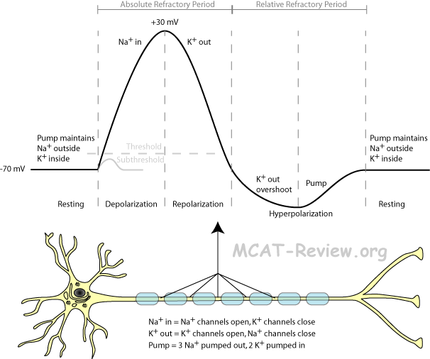
- Resting: cell at rest, sodium-potassium pump maintaining resting potential (-70 mV). Lots of sodium outside, lots of potassium inside. Ion channels closed so the established ion gradient won't leak.
- Depolarization: sodium channels open, positive sodium rushes inside, membrane potential shoots up to +30 mV. Lots of sodium inside, lots of potassium inside.
- Repolarization: potassium channels open, sodium channels close, positive potassium rushes outside, membrane potential drops back down. Lots of sodium inside, lots of potassium outside (opposite of the resting state).
- Hyperpolarization: potassium channels doesn't close fast enough, so the membrane potential actually drops below the resting potential for a bit.
- Refractory period: the sodium-potassium pump works to re-establish the original resting state (more potassium inside, sodium outside). Until this is done, the neuron can't generate another action potential. Absolute refractory period = from depolarization to the cell having re-established the original resting state. Relative refractory period = After hyperpolarization till resting state re-established.
- threshold, all-or-none
- When a stimulus (graded potential) depolarizes above a threshold value, an action potential will occur.
- Action potentials are all-or-none, meaning that if it occurs, all action potential have the same magnitude.
- One graded potential just barely makes the threshold value, another overshoots it a lot, but both will cause the same action potential.
- sodium-potassium pump
- 3 sodium out.
- 2 potassium in.
- net positive out.
- causes membrane to be more negative on the inside, hence negative membrane potential.
- Excitatory and inhibitory nerve fibers (summation, frequency of firing)
- Excitatory = stimulates an action potential to occur
- Excitatory synapse = receptor binding causes postsynaptic potential to be more positive (depolarization) = if it gets above threshold, action potential results.
- Inhibitory = inhibits an action potential from occuring.
- Inhibitory synapse = receptor binding causes postsynaptic potential to be more negative (hyperpolarization) = makes it more difficult to reach threshold.
- Summation = two or more nerves firing at the same time.
- Two subthreshold excitatory nerves firing at the same time can sum to reach the threshold.
- A threshold excitatory nerve and an inhibitory nerve firing at the same time, and the resultant signal won't reach the threshold.
- Frequency = Firing, then quickly firing again.
- If the first fire is subthreshold, fire again before the previous depolarization dies, and the new depolarization will be even higher than the first time.
Muscle Cell/Contractile
- Structural characteristics of striated, smooth, and cardiac muscle (old aamc topic)
- Striated = skeletal muscles, voluntary, has stripes, multiple nuclei shared within the same muscle fiber. Strong, but tire easily = shaped like long fibers.
- Smooth = visceral, involuntary muscles, no stripes, single nucleus per cell. Weak, but doesn't tire easily = shaped like almonds, tapered on both ends.
- Cardiac = heart muscles, involuntary, has stripes, single nucleus per cell, strong and doesn't tire easily = highly branched, shaped like fibers cross-linked to one another.
- Abundant mitochondria in red muscle cells (ATP source)
- Red muscle = high endurance, but slow.
- Aerobic respiration predominant.
- Many mitochondria because red muscles undergo aerobic respiration.
- Equipped to receive abundant oxygen supply: many capillaries, many myoglobin.
- High endurace, doesn't tire easily.
- White muscle = fast, but fatigue easily.
- Anaerobic respiration (glycolysis) predominant.
- Few mitochondria because white muscles undergo mainly glycolysis.
- Equipped for short bursts of glycolysis: stores high amounts of glycogen.
- Pink muscle = intermediate between red and white muscle.
- Organization of contractile elements (actin and myosin filaments, cross bridges, sliding filament model)

- Actin filament = thin filament = has troponin and tropomyosin on it.
- Myosin filament = thick filament = has myosin heads on it.
- Cross bridge = myosin head binds to actin.
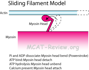
- Sliding filament model = Cross bridge forms, myosin head bends (power stroke), causes actin to move (slide) in the direction of the power stroke (toward the M line). When all the actin slide toward the M line like this, the muscle fiber contracts.
- Something counter-intuitive about the sliding filament model: ATP is not directly needed for the powerstroke. ATP binding is needed for detachment of myosin head to actin. ATP hydrolysis is needed for de-powerstroke (unbend myosin head).
- Rigor mortis = no ATP after a person dies, myosin heads can't detach after power stroke, muscle remain in contracted position.
- So what is troponin and tropomyosin there for? Ans: tropomyosin on actin blocks the myosin head from forming cross bridges. However, troponin moves tropomyosin out of the way at high Ca2+ levels (Ca2+ binds to troponin, and troponin moves tropomyosin).
- Calcium regulation of contraction, sarcoplasmic reticulum

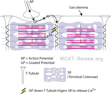
- Sarcoplasmic reticulum (SR) = smooth ER in muscle = stores calcium, releases them in response to AP.
- The SR is also called terminal cisternae where it meets T-tubules at the edges of the sarcomere.
- T-tubule = extension of the muscle cell membrane that runs deep into the cell, so that action potential can reach there.
- Muscle contraction:
- Nerve stimulates muscle.
- Action potential runs along muscle cell membrane.
- Goes deep into the muscle cell via T-tubules.
- Stimulates the SR (terminal cisternae) to release calcium.
- Calcium causes muscle to contract via the sliding filament mechanism.
- Sarcomeres ("I" and "A" bands, "M" and "Z" lines, "H" zone - General structure only)

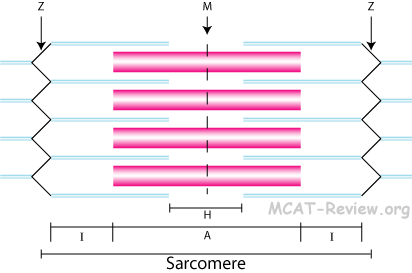
- I band = thinnest = thin filaments only = sides of the sarcomere.
- H zone = fattest = thick filaments only = center of the sarcomere, spans the M line.
- A band = contains both thick and thin filaments, center of the sarcomere spans the H zone.
- M line = line of myosin in the middle of the sarcomere, linked by accessory proteins.
- Z line = zigzag line on the sides of the sarcomere, connects the filaments of adjacent sarcomeres.
- mnemonics
- I = thin like the letter I.
- H = fat like the letter H.
- A = letter width in between I and H, so a mixture of thick and thin filaments.
- M = middle line = myosin (linked by accessory proteins).
- Z = zigzag line
- Moving from middle to the side of sarcomere = M HAIZ, the Muscle says HAIZ.
- Presence of troponin and tropomyosin
- Tropomyosin = long protein that spirals along actin, blocks myosin head from cross-linking.
- Troponin = binds tropomyosin, moves it out of the way when calcium is around.
Other specialized cell types
- Epithelial cells (cell types, simple epithelium, stratified epithelium)
- Squamous = flat.
- Cuboidal = cube.
- Columnar = column shaped.
- Simple epithelium = single cell layer = good for absorption, secretion, filtration, diffusion.
- Simple squamous: endothelium, capillary wall, alveolar wall.
- Simple cuboidal: gland ducts, kidney tubules.
- Simple columnar: stomach and gut.
- Stratified epithelium = two or more cell layers = good for protection against abrasion.
- Stratified squamous: skin.
- Stratified cuboidal/columnar: not common.
- Endothelial cells
- Endothelial cells = lines the inside of organs and blood vessels = simple squamous epithelium.
- Thin, single layer cells facilitate diffusion.
- Connective tissue cells (major cell types, fiber types, loose vs. dense, cartilage, extracellular matrix)
- Connective tissue structure = Cells + extracellular matrix.
- Cells: secrete the extracellular matrix (ground substance and fibers).
- Ground substance: glue that holds the matrix together.
- Fibers: mostly collagen, gives the matrix strength.
- Connective tissue cells and tissue types: bone, fat, tendons, ligaments, cartilage, blood.
- Osteoblasts make bone.
- Fibroblasts make connective tissue proper (fats, tendons, ligaments, beneath epithelia).
- Chondroblasts make cartilage.
- Hematopoietic stem cells make blood.
- Nomenclature:
- -blast = stem cell actively producing matrix.
- -cyte = mature cell, doing housekeeping.
- Fiber types:
- Collagen = the most common fiber type. Very strong. Present in large amounts in dense connective tissue.
- Elastic fibers = can stretch.
- Reticular fibers = can branch and form nets. Found in loose connective tissue.
- loose vs. dense
- Loose = loose fibers, lots of fluff (ground substance, cells) = anything that you don't associate with being fibrous = fat, paddings around organs.
- Dense = dense fibers predominantly collagen = genuinely fibrous, little fluff (ground substance, cells) = tendon, ligament.
- Cartilage = chondrocytes + matrix = elastic, flexible, used as padding in spinal discs, ends of bones, ear.
- Extracellular matrix = secreted by cells = ground substance (glue) and fibers.
|
|
